The language of ultrasound
The language of ultrasound is made up of descriptive words to try to form a picture in the reader’s mind. Ultrasound waves are formed in the transducer (the instrument the radiologist applies to the body), and reflect from tissue interfaces that they pass through back to the same transducer. So at every change in tissue type (eg. fat to kidney), some echoes pass through the interface and some are reflected. The reflected ultrasound waves form the image that we see on the screen.
Echogenicity
Because we are dealing with ultrasound waves, the descriptive terms are based in “echogenicity”, or the way the ultrasound wave is reflected back to the transducer. Each tissue type, such as liver, spleen or kidney, has a particular echogenicity in its normal state. In diseased states, the echogenicity of an organ can be altered, either more echogenic (hyperechoic) or less echogenic (hypoechoic) than usual. These observations can help the radiologist to categorize the type of disease process involved.
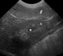
Echogenicity can be used to describe the tissue in relation to another tissue, or as compared to its normal state. For example, the mnemonic used to teach relative echogenicities is “my cat loves sunny places”. Taking the first letter from each word, the tissues go from hypoechoic to hyperechoic relative to each other. The renal medulla (inner portion of the kidney) is normally more hypoechoic than the renal cortex (outer portion of the kidney), which in turn is more hypoechoic than the liver, spleen and prostate. Echogenicity is not something that can be measured like ounces or centimeters, so using relative terms makes sense. In the image, the medulla of the kidney (*) is darker, or hypoechoic to the cortex of the kidney (^). The spleen (#) is hyperechoic to both of these tissues.

Echogenicity can also be used to mean “different than the normal echogenicity”. We usually try to compare a tissue to another normal tissue on the ultrasound screen, but it’s not always possible. The radiologist has a mental library of many images of normal tissue echogenicity, and will use that to compare the tissue on the screen as well.
What does it mean?
What does this mean for the animal being imaged? Ultrasound can give us very good information about problems within organs like the liver or spleen, such as picking up nodules (less than 4 cm diameter) or masses (greater than 4 cm diameter). It can also detect generalized changes in echogenicity of an organ.
For example, an enlarged, hyperechoic liver is brighter than the spleen. This can be caused by steroid administration, diabetes, or several other diseases. If there are nodules or masses that are hypoechoic to normal liver, hyperechoic, or mixed, we know that there are focal lesions but not what they are. The description helps a reader to “see” the lesion, but may not be specific to a particular disease. A hypoechoic lesion could be benign liver hyperplasia, which is very common in older dogs, or a cancerous nodule. Certain patterns, such as a “target” lesion, are more associated with cancer. If the diagnosis is unclear after ultrasound, a fine needle aspirate or biopsy might be recommended to determine what the nodule is.
Normal relationships in echogenicity of organs means that there is no apparent problem. If there are no nodules or masses, and the echogenicity of the organ is normal, we are more confident that there is no disease present. Ultrasound does have limitations in detecting disease; especially in the liver, spleen and kidneys, it may not detect subtle changes.
The take-home message
If your animal has an ultrasound examination, changes in echogenicity can help to pinpoint the organs that are affected. The pattern of change can suggest a range of problems, and is often not specific. Your radiologist and veterinarian will consult and determine the next step in diagnosing your animal’s illness. That may include blood tests, a fine needle aspirate or biopsy, or other diagnostic tests. Ultrasound is a very good tool to direct the diagnostic pathway.
Ultrasound terms:
- Hyperechoic – more echogenic (brighter) than normal
- Hypoechoic – less echogenic (darker) than normal
- Isoechoic – the same echogenicity as another tissue
Recent Comments