 A 14 year old dog came into our hospital last week with neurological signs, and was scheduled for an MR of the brain. He came to ultrasound for an abdominal scan to investigate the cranial organomegaly that the clinican palpated when he arrived. The first image that appeared on the ultrasound screen was a mass.
The mass was slightly heterogenous with a few cystic areas, and was located caudal to the liver. There was a small amount of free fluid in the abdomen. The mass measured 5×8 cm and filled the screen (figure 1). The next question is: where is it coming from? Liver or spleen?
It can be surprisingly difficult to determine the origin of a very large abdominal mass. They take up a lot of space, and can displace other organs altering your perception of normal anatomy. If the mass is pedunculated, the bulk of it can hide the connection to the other organ. Or the mass can involve the majority of the parenchyma making it difficult to find normal tissue. There are a few steps that can help you to make the diagnosis.
 Focus
Focus
Don’t spend all your time looking at the mass! It’s very easy to get impressed by the size and imaging characteristics, but you’ve already determined there is a mass. Focus on the origin.
Evaluate the liver
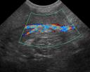
Look at all of the borders of the liver, from left to right under the xiphoid, and follow the lobes out to their periphery caudally. Can you find all of the serosal margins? Do any of them connect to the mass? If so, something that is very useful is to see if the blood supply is continuous from the liver to the mass. In our case (figure 2), there were portal veins connecting the liver on the left with the mass on the right of the image, so the diagnosis was a pedunculated hepatic mass.
Look for the spleen
Look for the spleen. If it’s a hepatic mass, the spleen could be pushed quite a bit more caudally or dorsally than you expect. If you aren’t getting a good view because of the mass, roll the animal into right lateral recumbency so you have good access to the left body wall. It can also be located under the ribs on the left side. If the mass is hepatic, as in our case, you will see a normal spleen from proximal to distal extremity (make sure you see the entire spleen). If the mass is splenic in origin, you may find a remnant of normal spleen that will increase your confidence in your diagnosis. If the mass has replaced the spleen, the fact that you found no normal spleen (and a normal liver) will also help.
Finally, an aspirate of the mass can help to determine its origin. Large masses can have necrotic centers, so use your Doppler to find a vascular place to sample. The periphery or the edge between normal and abnormal are usually good areas to try. Aspiration of the abdominal fluid for cytology can also provide a lot of clinical information.
Recent Comments