Infarction of the spleen or liver is a fairly rare event. But if the blood supply is reduced, a region, or the entire organ, can become infarcted. Infarction is necrosis resulting from a reduced blood supply. The main causes of infarction of the liver and spleen are malposition (torsion) and thrombosis.
Findings in splenic torsion
A torsed spleen is usually enlarged because of congestion. The arterial blood supply continues to enter the spleen, but is unable to exit through the collapsed veins that pass through the area of torsion. The spleen usually appears hypoechoic, with a “lacy” pattern of hyperechoic strands throughout (2). Focal patterns of splenic infarction have also been reported in splenic torsion, with hypoechoic or isoechoic splenic nodules. In early cases of torsion, the echogenicity of the splenic parenchyma may be normal. In more chronic cases, or septic spleens, you may see gas as bacteria invade the compromised tissue. The mesentery is hyperechoic around the enlarged spleen, and there is often peritoneal effusion as well. You may also be able to recognize that the spleen is in an abnormal position.
Doppler ultrasound
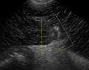
Since the blood supply to the spleen is the inciting problem in splenic infarction, evaluation of the arteries and veins with color and spectral Doppler ultrasound adds valuable information. Additional findings in dogs with splenic torsion include large splenic veins, echogenic venous thrombi, no visible thrombi, and lack of Doppler signal (3). The first image shows a lacy, hypoechoic, enlarged spleen with hyperechoic surrounding mesentery and absent Doppler flow in the splenic veins. For the veins and tissue itself, you need to make sure that your Doppler scale is set somewhere between -10 and 10 or so, since at higher detection speeds you could miss low flow. Power Doppler is a useful technique for evaluating low rates of blood flow in the splenic parenchyma.
Infarction vs. thrombosis
Another pattern that I have experienced is wedge-shaped areas of “lacy”, hypoechcoic appearance. These are usually seen in dogs with infarction by thrombotic events rather than splenic torsion. A recent study (4) also described a hyperechoic triangle on either side of the splenic veins in cases of splenic torsion, that is not found in infarction due to thrombosis. Differentiating torsion from thrombosis is important for deciding on surgical or medical treatment for the animal.
Liver infarction
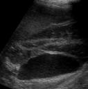
Similar to the spleen, liver infarction can be due to reduction of blood supply from mechanical causes (torsion, entrapment) or thrombosis.  Cases in the literature (5) have reported an enlarged liver lobe with hypoechoic, heterogeneous liver parenchyma and portal vessels with no blood flow and/or thrombi. Peritoneal effusion was a common finding. Liver lobe torsion can also be followed by abscessation. The second image shows a hypoerchoic liver lobe with a subtle lacy pattern and central portal vein with hyperechoic walls. This lobe is between the gall bladder (bottom of image) and a normal liver lobe (top of image). The hyperechoic striations are less apparent in the liver than in the spleen. The enlargement, lack of blood flow, peritoneal effusion and occasionally visible thrombi are other parallels. The mechanism of disease as well as the resulting ultrasonographic pathology are similar in hepatic and splenic infarction.
Findings in hepatic or splenic infarction
- enlargement
- malposition
- hypoechoic parenchyma with lacy pattern
- spleen – hypoechoic or isoechoic nodules, wedge-shaped lacy areas
- hyperechoic surrounding mesentery
- peritoneal effusion
- +/- intraparenchymal gas
- splenic torsion – triangular hyperechoic tissue surrounding veins
- Konde LJ, Wrigley RH, Lebel JL, et al. Sonographic and radiographic changes associated with splenic torsion in the dog. Veterinary Radiology 1989;30:41-45.
- Schelling CG, Wortman JA, Saunders HM. Ultrasonic detection of splenic necrosis in the dog. Veterinary Radiology 1988;29:227-233.
- Saunders HM, Neath PJ, Brockman DJ. B-mode and Doppler ultrasound imaging of the spleen with canine splenic torsion: a retrospective evaluation. Veterinary Radiology & Ultrasound 1998;39:349-353.
- Mai W. The Hilar Perivenous Hyperechoic Triangle as a Sign of Acute Splenic Torsion in Dogs. Veterinary Radiology & Ultrasound 2006;47:487-491.
- Schwartz SG, Mitchell SL, Keating JH, et al. Liver lobe torsion in dogs: 13 cases (1995-2004). J Am Vet Med Assoc 2006;228:242-247.
[…] Dr. Zwingenberger at Veterinary Radiology discusses hepatic and splenic infarction. […]