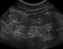
Ultrasound of linear foreign bodies can be more difficult than it seems. There are a couple of different imaging presentations depending on the size of the foreign body itself. I like to use radiographs to look for the pattern of linear foreign body, then confirm with ultrasound and look for complications.
With a linear foreign body, the proximal end is often wrapped around the base of the tongue (classic sewing thread in cats), or lodged in the pylorus. The trailing end of the string or rope then passes down the small intestine while remaining anchored. The intestine bunches up along this fixed foreign body, which causes the characteristic plication on radiographs. Plication is also visible on ultrasound, but can be more difficult to recognize until you’ve seen a few.
 The linear foreign body is taut, and so localizes on the mesenteric side of the intestine. The plication then folds the small intestine in a couple of different planes on the antimesenteric side. As a result, the plication can come in and out of your field of view on ultrasound (image 1). You can see some of the undulating folds, but not all unless you move the transducer to a different plane. The intestine is often not particularly dilated since fluid and gas can pass through the lumen.
The linear foreign body is often visible, but the appearance differs with the size and material. Thin linear foreign bodies, like string, tend to cause a hyperechoic line that can be seen in sections (Image 2). Because the linear foreign body is on the mesenteric border, and the plication is toward the antimesenteric border, the foreign body itself and the plication are often not visible in the same plane. Images 1 and 2 are from the same patient, illustrating this finding.
If the linear foreign body is large, like rope, it tends to cause a “clean” shadow artifact as it attenuates the ultrasound beam. You can take a look at a clean shadow here with a focal foreign body. Since these are often lodged in the pylorus, it pays to investigate the stomach thoroughly. Positional ultrasound can help to move gas away from the pylorus.
Of course, the surgeon will want to know the extent of intestinal involvement, so try to estimate the length of the small intestine involved. For example, “from the pylorus to the ileum, and I can’t rule out colon because feces is obscuring my view”. Other findings of note are free fluid and lymphadenopathy, which might indicate compromised bowel. If the bowel is leaky or perforated, there may be hyperechoic mesentery and free fluid in the region.
Diagnosing linear foreign body on ultrasound:
- Plication of bowel
- Visualization of linear foreign body (hyperechoic or clean shadowing)
- Free fluid
- Lymphadenopathy
- Hyperechoic mesentery
Recent Comments