Today’s case is a 9 year old male neutered mixed-breed dog who presented for acute paresis with tentative diagnosis of T3-L3 myelopathy. One lateral projection was obtained pre-myelogram, and two additional abdominal radiographs were obtained post-myelogram and hemilaminectomy.
Be sure to post your interpretations in the comments! Questions are always welcome.
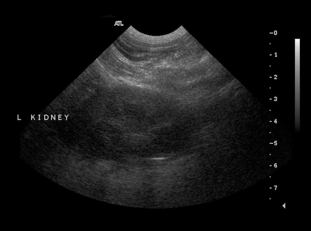
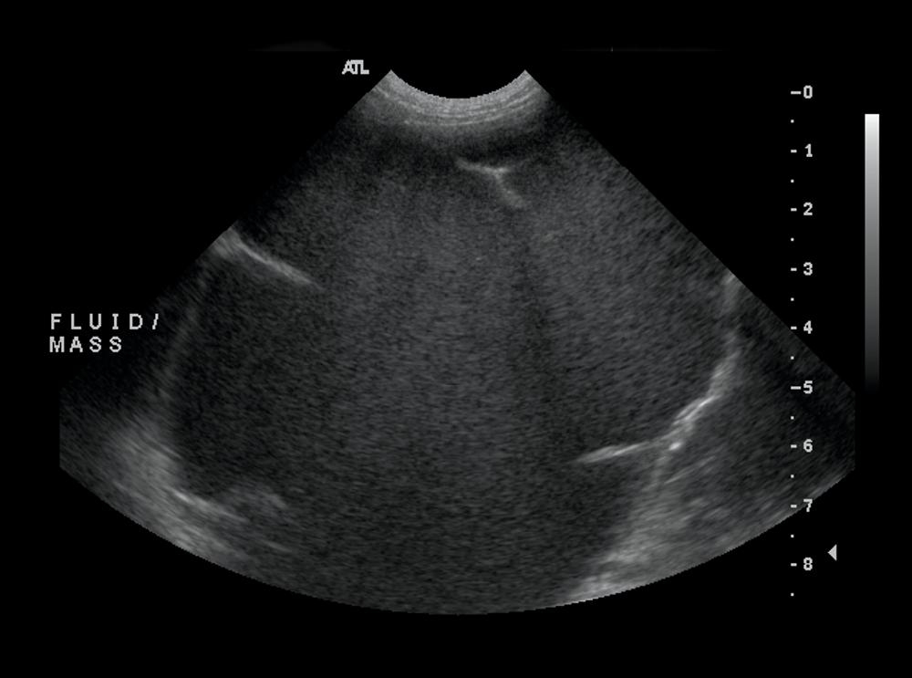
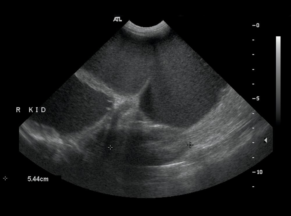
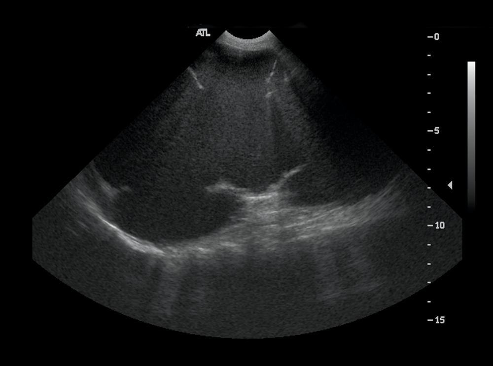
Ultrasound images
On ultrasound, the large, fluid-filled mass had multiple septae, which are characteristic of a hydronephrotic kidney and represent the connective tissue and arcuate arteries. The kidney was located in the central abdomen, so it was difficult to determine whether it was right or left. The left kidney is visible in the far field on the final image, so the hydronephrosis involved the right kidney.
Case originally posted on January 22, 2009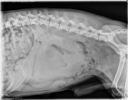
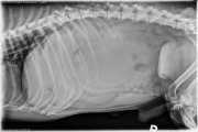
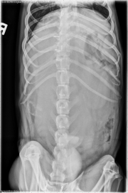
There is a large soft tissue mass within the mid to caudal abdomen causing compression and displacement of the adjacent gastrointestinal tract caudally and to the left, and the stomach cranially. There is a possible mild loss ofserosal details associated with it. In the 2nd and third rads there is contrast material in the UB. There are mineralized inter-vertebral discs- t13-L1, L1-L2. There is mineralized material in the right mid dorsal abdomen.
Possible sources of this mass can be a pedunculated liver mass, R kidny, GI tract, mesenteric LN. Ultrasound and FNAs will be helpful
That’s a good read. Where do you think that mineralized material in the right dorsal abdomen might be coming from? Does it alter the order of your differential diagnoses?