Abdominal ultrasound is a routine part of staging a new lymphoma patient. The liver and spleen are often involved, as well as intra-abdominal lymph nodes. So what do we expect to see if these organs are infiltrated with lymphoma?
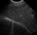
 The liver is very commonly infiltrated with neoplastic cells in multicentric lymphoma. The most common finding is an enlarged, hypoechoic liver, where the portal vessel walls are visible well out into the periphery (image 1). Occasionally the liver will look hyperechoic or isoechoic, and you may see a mass. Ultrasound is not very sensitive at detecting liver involvement with lymphoma. It often looks normal, even though it is diffusely infiltrated with lymphoma cells.
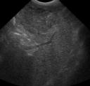
Ultrasound is much better at detecting splenic lymphoma. The spleen looks enlarged and hypoechoic, with a “swiss cheese” pattern. The hypoechoic nodules that make up the “holes” of the swiss chese are the infiltrated white pulp of the spleen. There may be a mass lesion as well, but diffuse involvement is most common (image 2).
There are usually multiple abnormal abdominal lymph nodes. Commonly seen lymph nodes like the cranial mesenteric and the medial iliac lymph nodes are easy to identify. Other chains of lymph nodes that are usually too small to see with ultrasound may become involved as well. It’s helpful to know the locations of some of these other groups like the periaortic and the splenic lymph nodes to be able to identify them. The lymph node enlargement can be very variable, with extremely enlarged nodes in one location, and mildly enlarged nodes in another. They tend to be hypoechoic and rounded. Their echotexture is usually uniform, but you may see mottled or nodular lymph nodes as well. There can also be a hyperechoic rim around the lymph nodes in the mesentery, which I think is inflammation or edema.
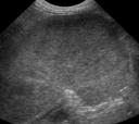
The spleen, liver and lymph nodes are the expected findings, but there is often something unexpected as well. Lymphoma can cause a renal mass (image 3), or the GI tract might be involved. It’s important to do a thorough examination to inform the clinician of any lesions that are out of the ordinary. Fine needle aspirate is an easy technique to confirm the diagnosis of lymphoma in these organs.
Lamb CR, Hartzband LE, Tidwell AS, et al. Ultrasonographic findings in hepatic and splenic lymphosarcoma in dogs and cats. Veterinary Radiology 1991;32:117-120.
[…] only thing I found about that spleen appearance is related to lymphoma (see third paragraph here : Abdominal ultrasound in canine lymphoma: expect the unexpected ). I would be so worried too and I am sorry I cannot give you better info. Others will very […]