Today’s case is a male Goose of unknown age and breed presented with weight loss and open mouth breathing. All the exotic and non-domestic specialists, post your comments!
Findings
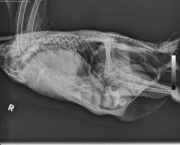
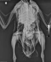
There is a large soft tissue mass in the cranial coelom displacing the trachea dorsally. In addition there is a smaller rounded soft tissue structure caudally and on the left adjacent to this mass measuring approximately 3 cm. There is moderate granular debris within the ventriculus. In addition there is a third rounded soft tissue structure just ventral to the pelvis.
Differential Diagnosis
Cranial celomic mass – neoplasia, granuloma, abscess
Diagnosis
- Cystic carcinoma, most likely of air sac origin
- Renal cyst
- Hepatic biliary carcinoma
Pathology
- Coelomic mass: air sac cystic carcinoma
- Air sacs: moderate generalized airsacculitis with regionally extensive epithelial hyperplasia, interstitial fibrosis, regional edema, and mixed inflammatory infiltration (heterophils and foamy macrophages, with occasional lymphocytes and plasma cells)
- Left kidney: perirenal cyst
- Liver: biliary adenocarcinoma
- Liver: mild to moderate multifocal portal infiltrates of lymphocytes, and diffuse hepatocellular vacuolization, focal adipocyte infiltration, capsular fibrosis with entrapped bile ducts
- Lung: multifocal anthracosis
- Testis: aspermatogenesis (seasonal breeder) and bilateral intraluminal foamy macrophages in the efferent ductules
- Mitral valve: moderate endocardiosis (gross diagnosis)
- Left stifle: mild chronic eburnation of articular surfaces and osteophyte formation (gross diagnosis)
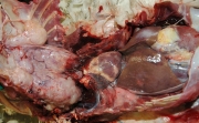
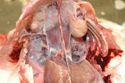
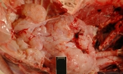
GROSS NECROPSY FINDINGS: This is an adult (>6 years old), 4.3 kg male goose in good nutritional state and postmortem condition but moderately dehydrated. The keel muscles are moderately atrophied. There is a large (14×8.5x8cm) mass occupying 70% of the thoracic cavity that is firmly attached to the ribs and the keel, compressing the lungs dorsally, and displacing the heart caudally. The mass is multilobulated, cystic, mottled tan to white, and friably gelatinous to semifirm, covered with thin semi-transparent capsule and supplied with a thin network of vessels. On cut surface, opaque fluid is emitted from the mass. There are 2 pedunculated, well demarcated, similar masses attached to the left caudo-lateral (3.5x2x1.5cm) and right caudo-lateral (~1cm in diameter) aspect of the ventral thoracic cavity that contain white pinpoint gritty material dispersed on cut surface. There is another similar mass attached to the cranial dorsal pole of the left kidney (2.2×1.5×1.2cm). Thoracic and coelomic air sacs are opaque and mildly thickened. Bilaterally, the cranioventral aspect of the lungs are 30% discolored brown red where they are compressed by the mass. The heart weighs 32.2g (0.75% of the body weight). The mitral valve is light tan, nodular and smooth. Measurements of the heart are as follows: right ventricular free wall (0.3cm), left ventricular free wall (1cm), and interventricular septum (1.1cm). The liver is subjectively enlarged, mottled light tan to pink, and extends well beyond the caudal extent of the keel and costal arches (weight 131.5g, 3% of the body weight). The external surface is roughened and multiple (>15) firm tan nodules are located on the surface as well as within the parenchyma (pin point to 0.8×0.7cm – largest nodule is in the right liver lobe). The nodule in the right liver lobe bulges on the cut surface and has irregular margins. The right testicle is slightly smaller than the left (~0.5cm vs 0.7cm in length). The mucosa of the intestines is coated with pink viscous material. Both the intestinal serosa and the mucosa of the caudal most extent of the ventriculus are diffusely discolored brown – red. There are irregular bony protrusions (osteophytes) that extend from the articular surface of the laterodistal femoral condyle (~0.3cm in diameter). The articular cartilage appears thin compared to the contralateral joint.
PRELIMINARY COMMENT: There is a large thoracic space occupying mass that likely contributed to the dyspnea and inappetance as described in the clinical history. A similar mass is attached to the cranial pole of the left kidney, and multiple nodules are within the liver.
References
- Raidal SR, Shearer PL, Butler R, and Monks D. Airsac cystadenocarcinomas in cockatoos. Australian Veterinary Journal. 84 96): 3213-216. 2007.
- Powers LV, Merrill CL, Degernes LA, Miller R, Latimer KS, and Barnes HJ. Axillary cystadenocarcinoma in a Moluccan cockatoo (Cacatua moluccensis). Avian Diseases. 42:408-412. 1998.
- Marshall K, Daniel G, Patton C, and Greenacre C. Humeral Air Sac Mucinous Adenocarcinoma in a Salmon-crested Cockatoo. Journal of Avian Medicine and Surgery. 18(3): 167-174. 2004.
Recent Comments