Cyclophosphamide has been known to induce a sterile, hemorrhagic cystitis in dogs when used in chemotherapy. As ultrasonographers, we are often asked to monitor the response to treatment of this condition. Today I’d like to show you some images of the progression of disease from acute through the healing process.
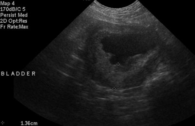
The first image is a transverse image of the bladder shows the thickened, irregular wall characteristic of cyclophosphamide-induced cystitis when first diagnosed. The mucosa is hyperechoic, which can be caused by hemorrhage. The bladder wall is approximagely 1.4 cm thick.
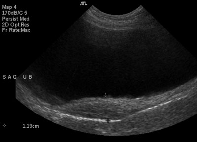
After two months of treatment, the majority of the bladder wall has returned to normal thickness and echogenicity (image 2). This sagittal image shows residual focal thickening in the dorsal aspect. The echogenicity is more normal, and the mucosal surface is more regular.
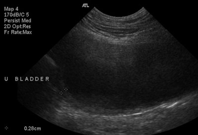
Finally, at 5 months (image 3, sagittal), the bladder wall is even and regular with very mild thickening at the cranial pole.
Does anyone else have experience imaging these? Post a comment here.


Hi, Allison, I saw two or three cases about cyclophosphamide induced hemorragic cystites, the ultrasound patterns is just like your. In one of the cases , only focal thickening at ventral part was identifid.
Great, thanks for your input!