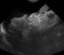
Chronic liver diseases cause damage to the normal liver parenchyma. Eventually, most of the hepatocytes are replaced with fibrous tissue and islands of nodular regeneration. When we see these cases in ultrasound, there are some characteristic changes to look for.
The liver is small and nodular
A small, nodular liver is very typical of cirrhosis. The liver can be very hard to see cranial to the stomach with the dog positioned in dorsal recumbency. A good approach is to use the intercostal spaces on the right and left sides to get cranial to the stomach and closer to the liver. In the first image, you can see the small liver lobes, with the contour distorted by a nodule (arrow).
Signs of portal hypertension
The fibrosis in the liver causes an increase in portal pressure. The portal system is a low-pressure system, with about 0-10 mm of mercury causing blood to flow from the intestines and splanchnic organs toward the liver. When the pressure in the liver rises above this, blood will flow retrograde along multiple extrahepatic shunts that open between the portal system and the caudal vena cava.
The things to look for in portal hypertension are ascites and the multiple extrahepatic shunts themselves. You can see the large amount of anechoic effusion in both images. In the second image, there are transverse and sagittal sections of vessels along the capsule of the kidney. These are multiple extrahepatic shunts, and they tend to form around the left kidney more so than the right. Color Doppler is a good way to demonstrate the vascularity in the region. In some cases, you may see reverse flow in the portal vein or one of its tributaries.
Things to look for:
- small, nodular liver
- ascites
- multiple extrahepatic portosystemic shunts
- hepatofugal flow in the portal vein


Recent Comments