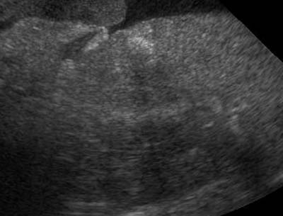
Renal infarcts are often incidental findings in animals with chronic renal disease. Infarcts are caused by thrombi that occlude a blood vessel in the kidney. Most of the time, they are chronic and mainly made up of fibrous tissue. The classic appearance is of a hyperechoic, wedge shaped area with … [Read more...]
Known Case Conference
KCC was another great session with interesting cases. Here are the highlights: Case 1 2 year old female neutered Rottweiler vomiting for 2 days. The abdomen in this dog had poor detail, and there were some very angular bowel loops and gas that might be free within the abdomen. Key point - … [Read more...]
Journal Club 10.24.07
Here are the latest articles in veterinary diagnostic imaging. Soler M, Murciano J, Latorre R, et al. Ultrasonographic, computed tomographic and magnetic resonance imaging anatomy of the normal canine stifle joint. The veterinary Journal 2007;174:351-361. Kofler J, Kneissl S, Malleczek D. MRI … [Read more...]
Ultrasound of arterial and venous thrombosis
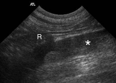
Thrombosis is a complication of many diseases in veterinary medicine. Heart disease, protein losing nephropathy and steroid therapy or hyperadrenocorticism can all predispose an animal to arterial or venous thrombi. Many of the systemic vessels involved are located in the abdomen and visible on … [Read more...]
Known Case Conference
This week we had a great mix of large and small animal cases. Case 1 - Mature dog with history of vomiting. There was a soft tissue opacity structure in the pylorus and duodenum on the v/d and left lateral projection. The descending duodenum was dilated and plicated in the right cranial abdomen. … [Read more...]
- « Previous Page
- 1
- …
- 160
- 161
- 162
- 163
- 164
- …
- 185
- Next Page »
Recent Comments