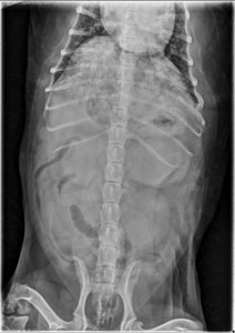
Localizing the mass If you see an obvious mass, the first step is to determine what quadrant of the abdomen it is in. Right cranial? Caudal midline? Mid-abdomen? When you narrow it down, you can make a mental list of the organs present in that area. For example, the right cranial quadrant contains … [Read more...]
Journal Club 4.23.07
Here are the latest journal articles. You may need a subscription to access them. They are also available on Citeulike. Join group VetRadiology and contribute your favorite articles! Lamb CR, Pfeiffer DU, Mantis P. Errors in Radiographic Interpretation Made by Veterinary Students. J Vet Med Educ … [Read more...]
Optimize radiographs for Powerpoint with Photoshop
Many of you, whether you are presenting a case for grand rounds, or for a meeting or more informal lecture, will be using images in your Powerpoint (or Keynote) presentations. It's hard to know what size, resolution and format to choose to end up with a quality image that is not so big that your … [Read more...]
What Are The Different Types Of Diaphragmatic Hernias? Here is another great question from the sophomore class about the different types of diapragmatic hernias. Hiatal hernia A Hiatal hernia happens when the cardia and fundus of the stomach pass cranially though the esophageal hiatus in the … [Read more...]
Splenic nodules
Splenic nodules or masses are extremely common findings when ultrasounding the canine abdomen. Nodules are small, circular abnormalities within the spleen that might be hyperechoic (brighter) or hypoechoic (darker) than the surrounding, normal spleen. Nodules are small (less than 4 cm diameter) and … [Read more...]
- « Previous Page
- 1
- …
- 173
- 174
- 175
- 176
- 177
- …
- 185
- Next Page »
Recent Comments