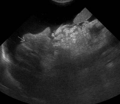
Chronic liver diseases cause damage to the normal liver parenchyma. Eventually, most of the hepatocytes are replaced with fibrous tissue and islands of nodular regeneration. When we see these cases in ultrasound, there are some characteristic changes to look for. The liver is small and nodular A … [Read more...]
How to hold the transducer
After teaching in the introductory ultrasound lab last week, I just want to post some quick tips on how to hold the transducer that several students found helpful. Hold the transducer like a pencil, not a flashlight. This gives your wrist the greatest range of motion for moving in different … [Read more...]
The ileocolic junction – where is it?
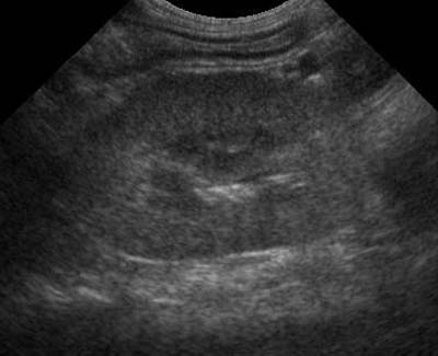
Everyone has their own pattern for performing an ultrasound exam. It helps to make sure you look at the major organs for signs of disease. In addition to the kidneys, gastrointestinal tract and pancreas, the ileocolic junction is a very important area to examine in cats. Cats are prone to … [Read more...]
Cyclophosphamide-induced cystitis
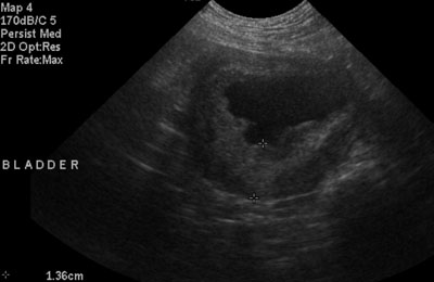
Cyclophosphamide has been known to induce a sterile, hemorrhagic cystitis in dogs when used in chemotherapy. As ultrasonographers, we are often asked to monitor the response to treatment of this condition. Today I'd like to show you some images of the progression of disease from acute through the … [Read more...]
Smallest ultrasound machine yet?
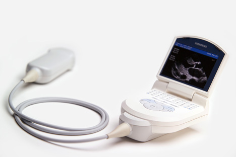
Medgadget reported on a Siemens handheld ultrasound machine that is the size of a Blackberry. The screen looks pretty small, but it would be interesting to see what it can do. Here is a link to the press release from Siemens, which states Additional applications include veterinarian medicine. … [Read more...]
- « Previous Page
- 1
- 2
- 3
- 4
- …
- 7
- Next Page »


Recent Comments